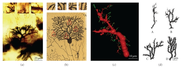
Figure 10.
(a) Shows Purkinje cell forming the dendritic branchlets, Cajal histological preparation, Golgi method, newborn cat. Insets: 1, terminal filopodia; 2, thin dendritic spines; and 3, dendritic spines contacting climbing fibers. (b) Purkinje cell, 15-day-old cat. Insets show filopodia and thin dendritic spines, Cajal scientific drawing [23, Figure 2]. (c) Purkinje cell, apical branch, three-dimensional reconstruction showing dendritic filopodia and protospines (green) and few dendritic spines (red), newborn cat. (d) Purkinje cell, dendritic filopodia, protospines and dendritic spines, rat of 5 days (A), 10 days (B), 15 days (C), and 30 days (D); scientific drawing of Berry and Bradley [24].

