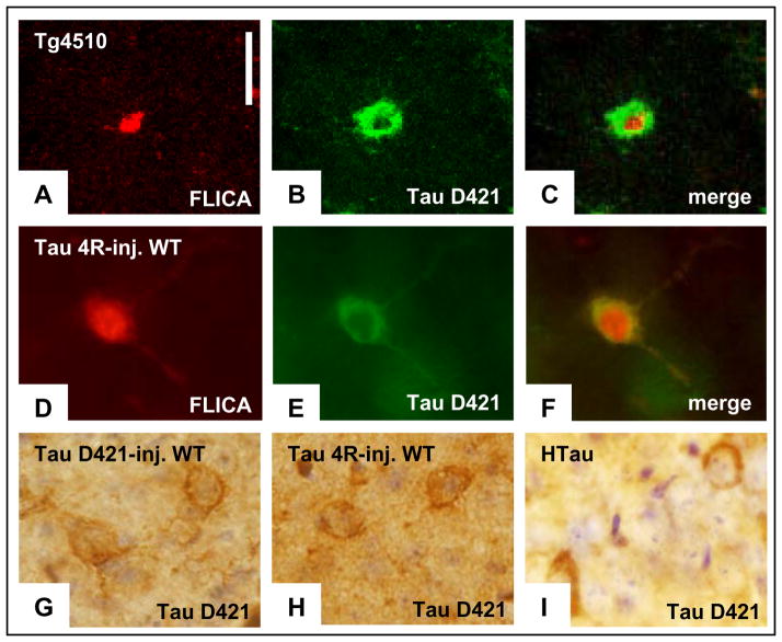
Figure 3. Caspase activation in Tg4510 correlates with the presence of truncated forms of tau, which appears in multiple models of tauopathy.
a, Pan-caspase fluorescent indicator applied in vivo can be detected in post-mortem tissue (red). b, c, Tau truncated at Asp 421 (labelled with Alexa Fluor 488, b) was detected using immunochemistry in the same cells that were positive for caspase activation (merged in c). All caspase-positive neurons were also positive for tau-D421. d, In wild-type (WT) animals, injection of AAV encoding tau-4R resulted in caspase activation detected in post-mortem sections by the persistent fluorescent indicator. e, f, Presence of truncated tau (labelled with Alexa Fluor 488, e) was detected in the same neuronal cell bodies using immunochemistry (merged in f). i, Tau-D421-positive neurons were observed in the hippocampus of 14-month-old hTau animals. h–g, The immunodetection of tau-D421 in neurons of the tau-4R virus-infected (h) and hTau (i) brains seems very comparable to signal detected in the neurons infected by the AAV encoding tau-D421 (g). Scale bars, 10 μm.

