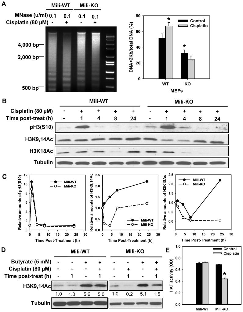
Fig. 1.
Mili is required for cisplatin-induced chromatin relaxation and histone acetylation. (A) Mili-WT and KO MEFs were treated with cisplatin for 1 h, further cultured in drug-free medium for another 1 h. The nuclei were isolated and subjected to MNase treatment. The genomic DNA was then isolated and resolved in agarose gel. The intensity of each lane was scanned and the ratio of DNA<2Kb to total DNA was calculated. The experiment was repeated three times. Bars represent SD. *: p < 0.05 compared with WT control. (B) Mili-WT and KO MEFs were treated with cisplatin for 1 h, further cultured in drug-free medium for various time periods. The phosphorylated and acetylated histone H3 was detected using Western blotting. (C) Quantitative analysis of modified histone H3 from the blot in (B), normalized to Tubulin. (D) MIli-WT and Mili-KO cells were pre-treated with Sodium butyrate for 0.5 h, before being treated with cisplatin for 1 h in the presence of butyrate. The cells were then cultured in the presence of butyrate for another 1 h. Acetylated H3 was detected using Western blotting. The band intensities were quantitatively assessed by scanning programs, and normalized to Tubulin levels. (E) Mili-WT and KO MEFs were treated with cisplatin for 1 h, further cultured in drug-free medium for another 2 h. The nuclear extract was prepared, the HAT activity was assayed and expressed with OD values. The HAT activity is an average of three independent repeats. Bars represent SD. *: p < 0.01 compared with control.

