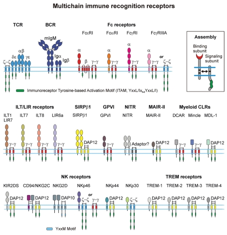
Figure 2.
Schematic presentation of the MIRRs expressed on many different immune cells including T and B cells, natural killer cells, mast cells, macrophages, basophils, neutrophils, eosinophils, dendritic cells and platelets. The inset shows MIRR assembly. The extracellular recognition domains and intracellular ITAM-containing signaling domains are located on separate subunits bound together by noncovalent transmembrane interactions (solid arrow). ITAMs/YxxM are shown by green. Curved lines depict intrinsic disorder of the cytoplasmic domains of MIRR signaling subunits. Abbreviations: BCR, B cell receptor; CLR, C-type lectin receptor; DAP-10 and DAP-12, DNAX adapter proteins of 10 and 12 kD, respectively; DCAR, dendritic cell immunoactivating receptor; GPVI, glycoprotein VI; ILT, Ig-like transcript; KIR, killer cell Ig-like receptor; LIR, leukocyte Ig-like receptor; MAIR-II, myeloid-associated Ig-like receptor; MDL-1, myeloid DAP12-associating lectin 1; NITR, novel immune-type receptor; NK, natural killer cells; SIRP, signal regulatory protein; TCR, T cell receptor; TREM receptors, triggering receptors expressed on myeloid cells.

