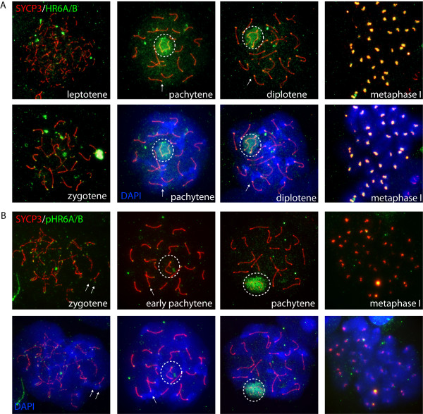
Figure 1.
HR6A/B localizes to centromeric chromatin in early pachytene. A, B: Immunostaining of spread spermatocyte nuclei for nonphosphorylated HR6A/B (green, A), phosphorylated HR6A/B (green, B), and SYCP3 (red A, B), The merge with DAPI (blue) staining for DNA is shown for a selection of nuclei. The centromeric ends of the SCs can be identified based on the more intense DAPI staining. Arrows indicate centromeric ends that are positive for phosphorylated and non-phosphorylated HR6A/B. The XY body is encircled. During leptotene and zygotene, both phosphorylated and nonphosphorylated HR6A/B accumulate as foci in the nucleus and on the developing SCs. Some centromeric and telomeric ends of the SCs also are enriched for phosphorylated and non-phosphorylated HR6A/B. During early pachytene, the XY body is not enriched for phosphorylated HR6A/B. At this stage, a clear accumulation of phosphorylated HR6A/B is observed on the centromeric ends of the SC. During late pachytene, this staining is lost, and phosphorylated and non-phosphorylated HR6A/B are prominent on the XY body. At metaphase of the first meiotic division, HR6A/B is highly enriched at centromeres, but not phosphorylated.

