Figure 8.
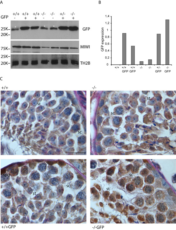
Enhanced X-linked GFP expression in Hr6b knockout testes. A, Western blot analyses of X-GFP, MIWI and TH2B expression in protein extracts derived from wild type (+/+) Hr6b+/- (+/-) and Hr6b-/- (-/-) mouse testes with or without the X-linked GFP transgene (GFP). MIWI (expressed in spermatocytes and early round spermatids) and TH2B (expressed from the spermatogonia stage onwards) are shown as controls. The white asterisks indicate a nonspecific band migrating slightly faster than the GFP band. B, quantification of the Western blot data shown in A. GFP signal was quantified using Image J software. The intensity of the background band in lane 1 was subtracted from the GFP signal in all lanes. Subsequently, the signal was normalized to the TH2B signal, and is shown on the Y-axis of the graph. C, immunohistochemical analyses of GFP expression in testis from mice that are wild type (+/+), wild type carrying X-GFP (+/+GFP), Hr6b knockout (-/-), or Hr6b knockout carrying X-GFP (-/-GFP). Al low level of nonspecific background brown staining is observed in the +/+ and -/- testes. Specific brown GFP staining is observed in spermatogonia (spg) in wild type mice carrying the X-GFP gene, but also in pachytene spermatocytes (spc) and round spermatids (spt) on the Hr6b knockout background. Size bar indicates 20 μm.

