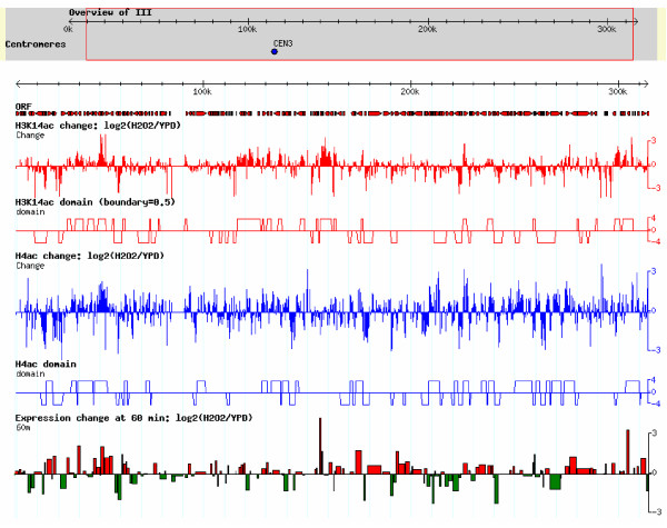
Figure 6.
Coacetylation domains in chromosome III. The figure was generated by GBrowse (which is provided by GMOD: Genetic Model Organism Database; http://gmod.org). The tool can present the gene information in the genome and the user's data in the corresponding genomic locations. The top gray region in figure is the overview of chromosome III, and the coordinate indicates the location of the chromosome. The red arrows under "ORF" showed the locations and transcribed directions of genes. The red vertical line under H3K14ac change: log2(H2O2/YPD) and the blue line under H4ac change:log2(H2O2/YPD), respectively, show the change of H3K14ac and H4ac under H2O2-stress conditions in each probe. The red rectangles under H3K14ac domain (boundary 0.5) and the blue rectangles under H4ac domain indicate the domains with similar changes of H3K14ac and H4ac, respectively. The bottom subfigure is gene expression. The height and width of the rectangle indicate the degree of gene expression change and ORF length, respectively, and the color indicates up-regulated (red) or down-regulated (green).

