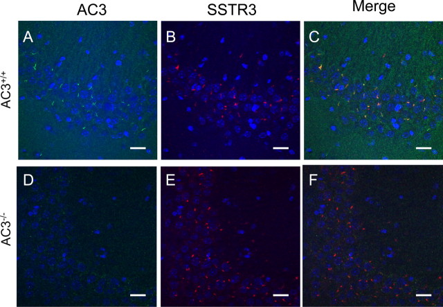
Figure 2.
Hippocampal neurons in AC3−/− mice express primary cilia. A–C, Representative images of AC3-immunoreactive cilia in the CA3 region of the hippocampus in AC3+/+ mice labeled with antibodies against AC3 (green) (A), SSTR3 (red) (B), and colocalization of AC3 and SSTR3 (merge) (C). Scale bar, 20 μm. D–F, Representative images of immunoreactive cilia in the CA3 region of the hippocampus in AC3−/− mice labeled with antibodies against AC3 (green) (D), SSTR3 (red) (E), and colocalization of AC3 and SSTR3 (merge) (F). Scale bar, 20 μm. n = 4 for each genotype. Blue, Hoechst nuclear staining.

