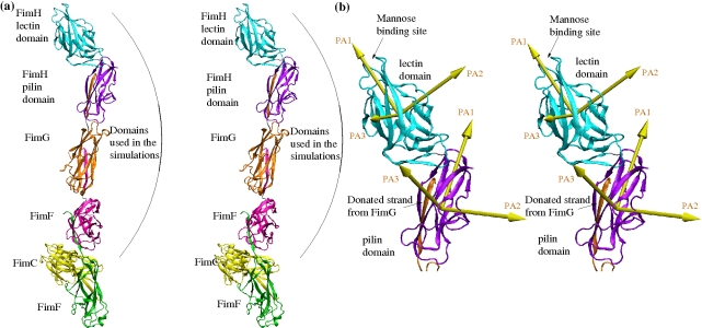
Figure 1. Crystallographic structures.
(a) Entire fimbrial tip (stereoview). Subunits are distinguished using different colors. A subunit consists of a polypeptide chain, whereas a domain is defined as the globular part of a subunit and (with the exception of the FimH lectin domain) a donated strand of the neighboring subunit (see “Materials and Methods” for the exact definition of start and end residues of domains). The FimH subunit consists of two domains, which are also distinguished with different colors: cyan for the lectin and purple for the pilin domain, respectively, except for the donated strand of FimG, which is colored orange as the subunit it belongs to. The two FimF subunits are colored magenta and green, respectively. The chaperone protein FimC is in yellow. Residues that are located between domains define linker chains and their backbone is colored in silver. The arrows in yellow represent the principal axes (PA1, PA2, and PA3) of each domain (see “Materials and Methods” for the determination of principal axes). They are used to calculate hinge and twist angles between adjacent domains (Figures S3, S4, S5d,e). (b) FimH subunit with donated FimG strand (stereoview). The principal axes of the FimH subunit are labeled in brown.

