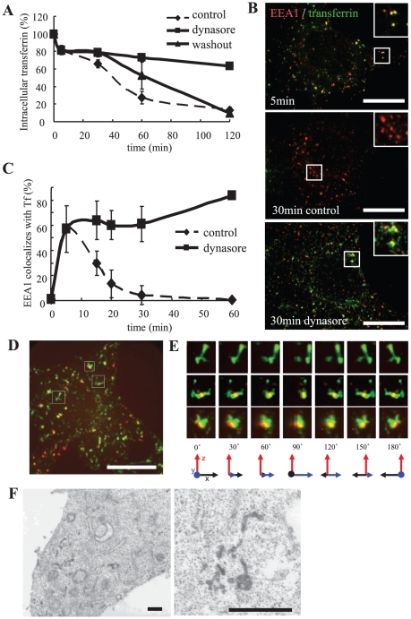
Figure 2. Dynasore inhibits transferrin recycling in early endosomes.
A, Cells were incubated with biotin-conjugated transferrin on ice for 1 h, rinsed, and transferred to 37°C. At each time point, cells were lysed and intracellular transferrin was measured by ELISA. In the ELISA assay, the cell lysates bound to plates coated with goat anti-transferrin antibodies (EY Laboratories, San Mateo, CA) and were detected with streptavidin-HRP. B, HeLa cells were incubated with Alexa 488-transferrin (Molecular Probes) on ice for 1 h, and then processed as in A. After 5 min, the media was changed to medium containing DMSO (control) or dynasore. At the time indicated, the cells were fixed and stained for EEA1 (BD Transduction Laboratories, San Jose, CA). Scale bar, 20 µm. C, Colocalization of EEA1 and transferrin was measured and is represented mean ± S.D. N = 20 cells. D, HeLa cells were bound with Alexa 555-EGF (Molecular Probes) and Alexa 488-transferrin on ice for 1 h. After rinsing, the cells were incubated at 37°C for 5 min and dynasore was added. Thirty minutes after internalization, the cells were fixed and observed. Scale bar, 20 µm. Pictures were processed for 3D reconstruction using MetaMorph software (E). F, Cells were bound to HRP-transferrin and treated for 30 min as in D. The cells were then incubated in a DAB-containing solution on ice for 30 min, fixed, and processed for electron microscopy. Scale bar, 500 nm.

