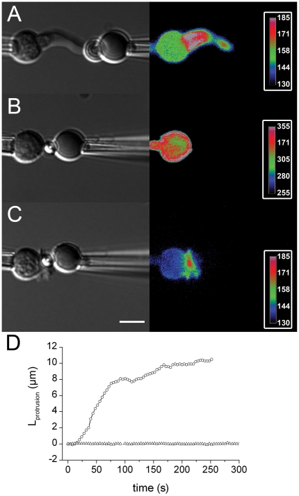
Figure 6. Cytoskeleton remodeling in T cells interacting with Abs-coated beads on the force probe.
T cells were transfected with LifeAct-mCherry (left : DIC, right : color code mCherry). (A) Anti-CD3 coated bead. (see Movie S6. (B) Anti-CD18 coated bead. (C) Anti-CD3+anti-CD18 coated bead. (D) Lprotrusion versus time for one representative experiment with a latrunculin A treated T cell (open triangles) or a non-treated T cell (open circles).

