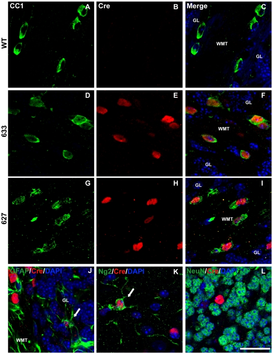
Figure 3. Cre-recombinase is predominantly expressed in oligodendrocytes of P28 Plp-Cre mouse cerebellum.
A–I. Cerebellar sections were double-stained with antibodies specific to CC-1 as a marker for mature oligodendrocytes (green), Cre (red) and counterstained with DAPI. Cre was not detected in WT cerebellar sections (B). Sections from transgenic mice of both F633 (D–F) and F627 (G–H) lines exhibited a similar pattern of Cre expression. High levels of Cre recombinase protein was detected in the CC1-positive oligodendrocytes. J. Cerebellar section stained for GFAP (green) as a marker for astrocytes, Cre (red) and counterstained with DAPI. A small number of weakly stained Cre-positive cells co-labeled with GFAP (arrow). K. Representative example within the molecular layer of cerebellar section stained for Ng2 (green) as a marker for OPCs, Cre (red) and counterstained with DAPI. A small number of weakly stained Cre-positive cells co-labeled with Ng2 (arrow). L. Representative example within granular layer of cerebellar section stained for NeuN (green) as a neuronal marker, Cre (red) and counterstained with DAPI. There were no NeuN positive cells co-labeled with Cre. GL = granular layer, WMT = white matter tract. Scale bar = 20 µm.

