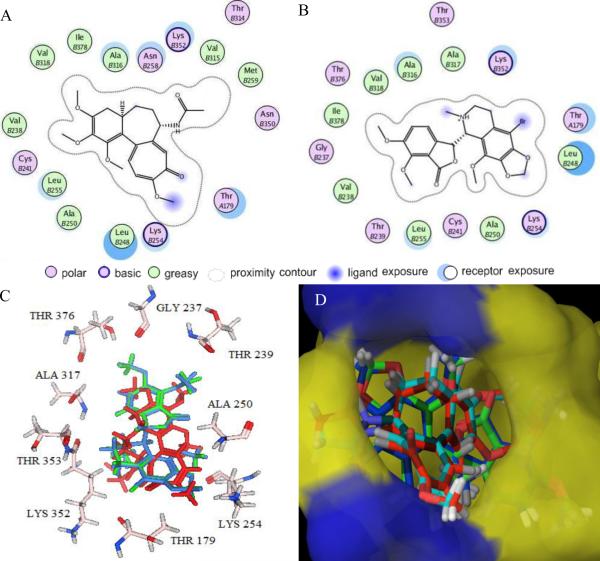
Figure 4. Results of Glide XP docking of colchicine, noscapine, and Br-noscapine into the colchicine binding site of tubulin.
2D representations of the binding site with amino acid residues involved in the interaction of (A) colchicine and (B) Br-noscapine reveal identical sets of amino acids. (C) 3D representations of the binding site amino acids within a 4Å distances from superimposed conformations of docked noscapine (blue) and Br-noscapine (green). The co-crystal structure of colchicine is represented in red color. Some of the amino acids (Leu248, Cys241, Val318, Ala316 and Val238) are not shown for better clarity. (D) Overlapped docking poses of colchicine, noscapine, and Br-noscapine obtained form Glide docking onto co-crystal of colchicine within the colchicine binding site of tubulin. Tubulin is represented as Macromodel surface according to residue charge (electropositive charge, blue; neutral, yellow) as implemented in Maestro.

