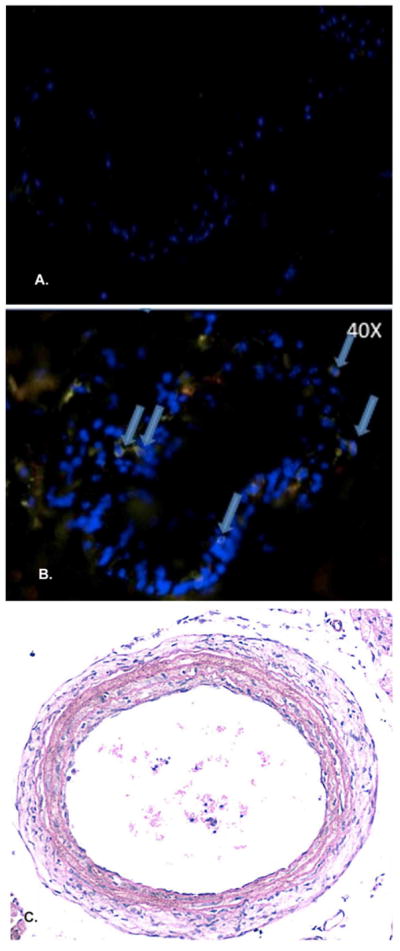
Figure 3. Lack of significant inflammation in the injured NOD.scid arteries.

In the setting of immunodeficiency, the NOD.scid shows the presence of few T cells (arrows) in the injured arterial wall at 7 days post-ligation (A [10X], B [40X]). Importantly, the NOD.scid demonstrates no appreciable neoinitimal formation at the 28 day time point (C).
