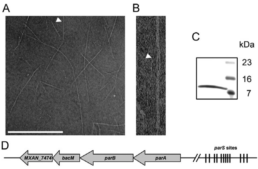
Fig. 1. Characterization of isolated BacM fibres and bacM-containing operon.
A. Electron micrograph of isolated purified BacM fibres. The arrowhead points to an individual fibre of about 3 nm width as it exits a fibre bundle. Bar represents 500 nm.
B. Magnified picture of the individual fibre marked in (A).
C. SDS-PAGE gel of these purified fibres shows a single band of about 11 kDa (right lane, MW marker).
D. Schematic of the operon containing bacM. The four open reading frames encode ATPase ParA (MXAN_7477), DNA-binding protein ParB (MXAN_7476), BacM (MXAN_7475) and putative lipoprotein MXAN_7474. Between 4.4 and 6.6 kilo base pairs (kbp) upstream of parA are twelve parS sites implicated in ParB binding. The regions comprising 0.5 kbp downstream and 8.7 kbp upstream of the operon do not encode known proteins. Note that the schematic is not drawn to scale.

