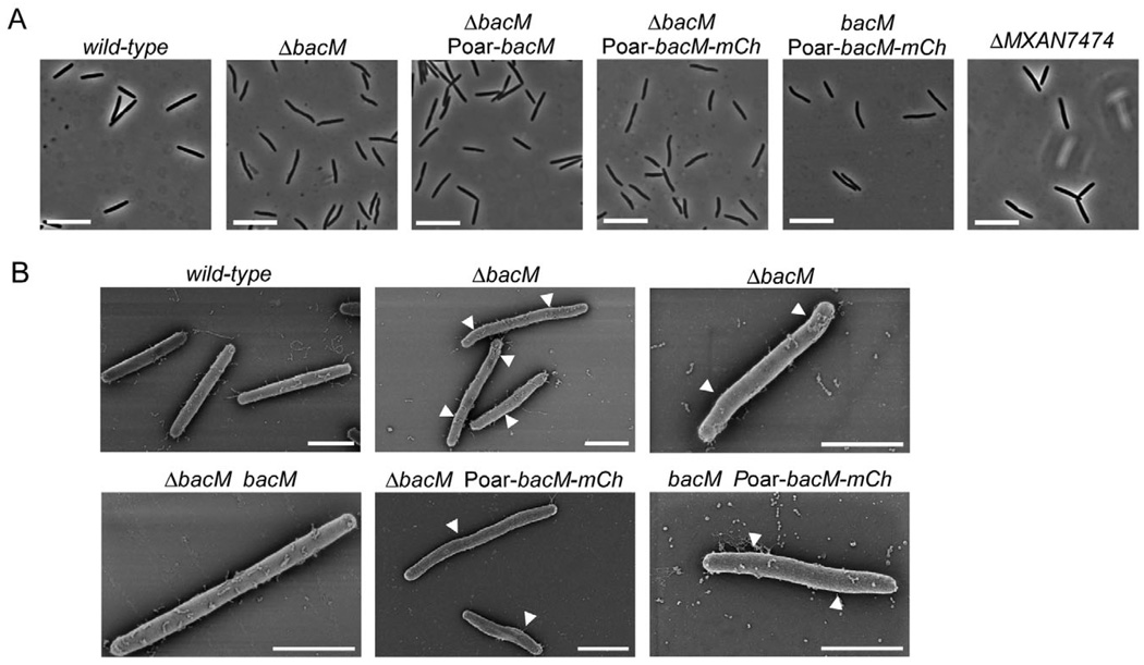
Fig. 2. Cell morphology of bactofilin mutants. Relevant genotypes are indicated.
A. Phase-contrast light micrographs of cells from liquid CTT cultures in mid-logarithmic growth phase. Bars represent 10 µm.
B. Scanning electron micrographs of cells grown as in (A). White arrowheads mark kinks along the cell body. Bars represent 2 µm. The following strains were analysed: DK1622 (wild-type), EH301 (ΔbacM), EH344 (ΔbacM Poar-bacM), EH364 (ΔbacM Poar-bacM-mCh), EH362 (bacM Poar-bacM-mCh), EH332 (ΔMXAN_7474), EH309 (ΔbacM bacM).

