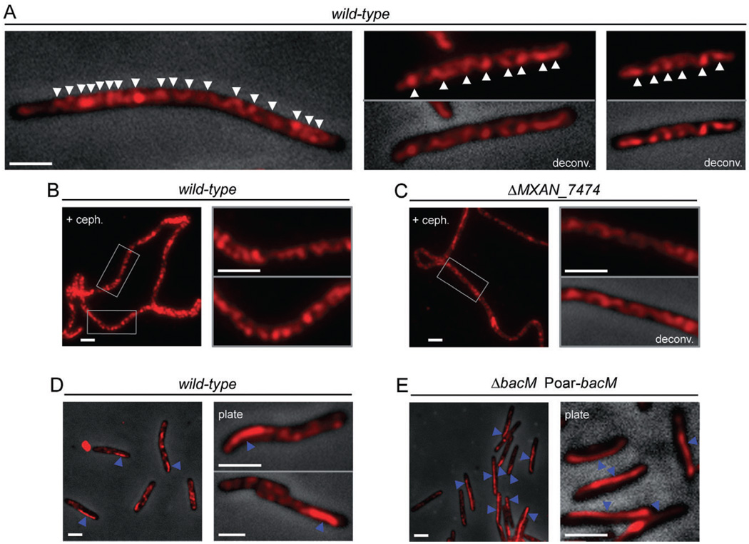
Fig. 4.
Subcellular localization of BacM. Immunofluorescence micrographs of M. xanthus cells treated with anti-BacM antibody and Alexa594-coupled anti-rabbit IgG as secondary antibody. Shown are either only the fluorescent signals or their overlays onto phase-contrast pictures. Fluorescent signals were deconvoluted when indicated (deconv.). Cells were grown in CTT medium to mid-logarithmic growth phase, or, when indicated, collected from CTT agar 8 days after plating (plate).
A. Cells of wild-type strain DK1622. White arrowheads indicate apparent turns of intracellular helix-like cables of BacM (variable spacing, generally between 0.5 and 1.0 µm).
B. Elongated DK1622 cells after growth for 14 h with cephalexin are shown at lower magnification and boxed regions are magnified in the adjacent pictures.
C. Cells deleted in MXAN_7474 (strain EH332) were treated with cephalexin and displayed as described for wild-type.
D. In addition to the cables, approximately 25% of DK1622 cells contain a mostly lateral polar rod-like structure (blue arrowheads).
E. Cells (strain EH344) deleted in bacM and subsequently complemented with the bacM gene under control of the oar promoter (Poar) generally contain lateral rod-like structures that can extend through the whole cell (blue arrowheads). White bars represent 2 µm.

