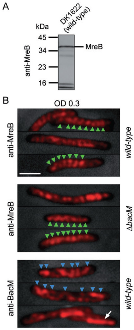
Fig. 6. MreB and BacM have different cellular localization patterns.
A. Immunoblot of DK1622 total proteins with anti-MreB antibody. A major band is present at approximately 36.5 kDa, the MW of M. xanthus MreB.
B. Anti-MreB fluorescence micrographs of wild-type (DK1622) and ΔbacM (EH301) cells from CTT cultures of OD 0.30. Bars represent 2 mm. After fixation, the cells were treated with either anti-MreB or anti-BacM primary and fluorophore-coupled secondary antibodies. Fluorescence signals are shown on top of phase-contrast pictures of the cells. In a selection of cells, green arrowheads mark the regular banding pattern present after staining with anti-MreB. When staining wild-type cells from the same batch with anti-BacM antibody, the observed pattern was less regular (blue arrowheads), and in about 25% of the cells, a lateral rod-like region near one of the cell poles (white arrow) showed stronger fluorescence compared with the rest of the cell.

