
Fig.1.
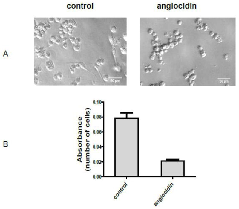
Morphological appearance and growth of angiocidin-treated MB-231 cells. MDA-MB-231 cells were seeded in 0.1% BSA DMEM without recombinant angiocidin and incubated at 37° C for sixteen hours. After sixteen hours the cells were photographed. The number of adherent, spread cells resembling the morphology of normal MB-231 cells in four representative fields was counted. The percentage of adherent, spindle-shaped cells was determined to be 36 out of 225, or 16% (panel A). Cell proliferation was determined in a 96 well plate using the Alamar blue proliferation assay under the same culture conditions (Panel B). Experiments were repeated three times and the results of a representative experiment are shown in the figure.