
Fig. 3.
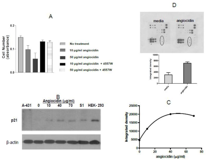
Angiocidin inhibits proliferation of MB-231 breast cancer cells through activation of EGFR and up-regulation of p21waf1. Panel A- Cells were grown in DMEM media containing 10% serum and 10 ng/ml EGF either alone or treated with 10 μg/ml or 50 μg/ml angiocidin in the presence or absence of 10 μg/ml EGFR inhibitor 4557W and proliferation was measured after 24 hours using the Alamar blue proliferation assay. Panel B- Cells were grown in DMEM media containing 2% serum for 24 hours in six well plates and then treated with various concentrations of angiocidin for an additional 24 hours, harvested and blotted for p21waf1. Cell lysate from A-431 was used as the negative control and cell lysate from HEK-293 cells was the positive control. Panel C- p21waf1 positive bands in Panel B quantitated by densitometry using Image J software. Panel D- EGFR phosphoarray results (top-stained array, bottom- spots encircled in the stained array were quantitated by densitometric analysis of the spots using Image J software ). Experiments were repeated two times and the results of a representative experiment are shown in the figure.