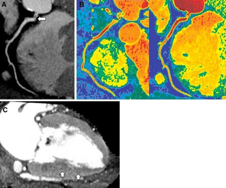
Fig. 7.
Coronary and myocardial images during adenosine stress in a 55-year-old male with severe stenosis of the right coronary artery. a Curved multiplanar reformatted image at 120 kV shows severe ostial stenosis of the right coronary artery (arrow). b Colored coronary images demonstrate higher attenuation within the coronary artery on the 120 kV images (left) than on the iodine map (right). The average attenuation for the right coronary artery was 338 HU on the 120 kV images and 318 HU on the iodine map. The CER value was 0.82 on the 120 kV images and 0.77 on the iodine map. The color scale used is the same as that in Fig. 4b. c Vertical long-axis slice (myocardial image) reconstructed from the same data as the 120 kV coronary angiogram shows a region of subendocardial hypo-enhancement in the inferior wall (arrows), suggestive of myocardial ischemia

