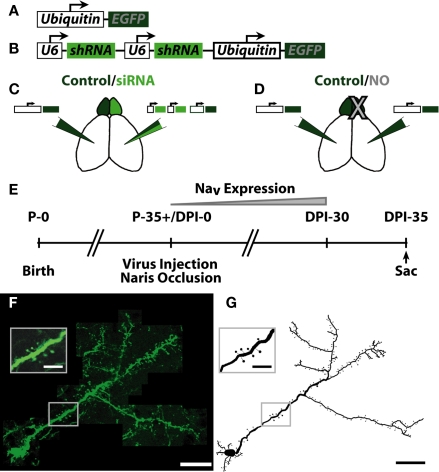
Figure 1.
Summary of experimental conditions and injection paradigm. (A) Schematic of EGFP coding lentivirus. EGFP was expressed downstream of the ubiquitin promoter. (B) Schematic of siRNA + EGFP coding lentivirus. Two U6 promoters, each expressing one shRNA against voltage gated sodium channel 1.1–1.3 (NaV1.1–1.3) expression were followed by the ubiquitin promoter expressing EGFP. Each group received the following manipulations: (C) – Control EGFP lentivirus [Control, see (A)] in one hemisphere, siRNA sodium channel knock-down lentivirus [siRNA, see (B)] opposite hemisphere. (D) – Control EGFP lentivirus in both hemispheres, naris occlusion (NO) in one hemisphere. (E) Experimental timeframe. Mice were allowed to survive for 35 days post-injection (DPI)/naris occlusion before sacrifice, allowing for expression of NaV in progenitor cells born the day of injection (DPI-0). (F) Example neuron from individual slices of an image stack. Scale bar, 100 μm. Inset shows resolution of apical dendrite spines. Scale bar, 25 μm. (G) Example ABN reconstruction from images in (F). Scale bar, 100 μm. Inset shows reconstruction of inset in (F). Scale bar, 25 μm.

