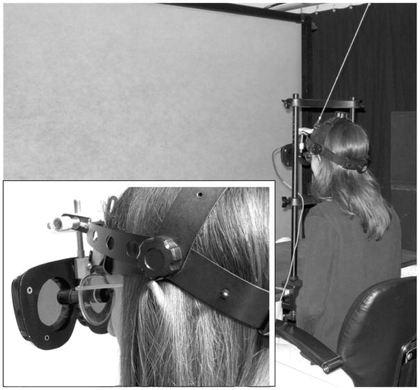
Figure 3.
The apparatus used in this study. Participants wore a modified indirect ophthalmoscope headband with the shutter lenses suspended forward and sat 1m from the screen where the fixation target and stimuli were presented. Inset (lower left) shows details of how the shutter lenses were suspended (the chin rest support bar was removed for the photo of the inset).

