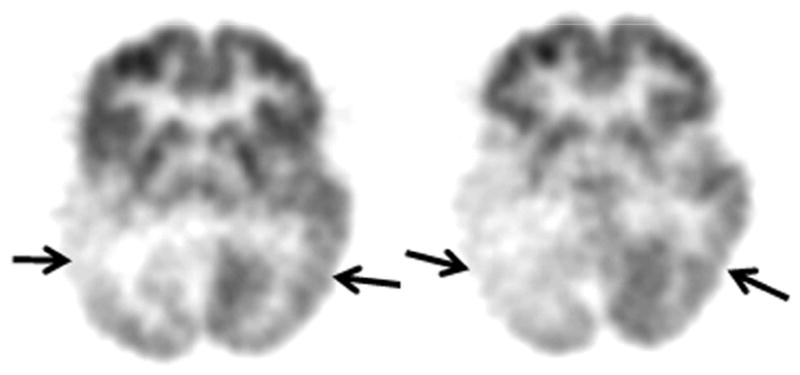
Figure 2.

Axial image planes of the glucose PET scan of patient # 5, whose developmental impairment was mild/moderate. However, he developed autistic behavioral features. His PET scan demonstrated bilateral (right more severe than left) temporal hypometabolism (solid arrows) as well as bilateral parietal and right occipital hypometabolism.
