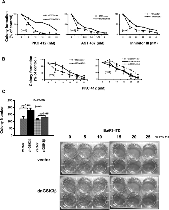
Figure 4.

GSK-3β inhibitions attenuate the response of Ba/F3-FLT3-ITD cells to the FLT3 kinase inhibitors on colony formation in a Wnt/β-catenin pathway–dependent manner. (A) Stable expression of dnGSK-3β in Ba/F3-FLT3-ITD cells decreases the potencies of the FLT3 kinase inhibitors on colony formation. Both Ba/F3-FLT3-ITD-vector and Ba/F3-FLT3-ITD–dnGSK-3β cells were seeded at a density of 500 cells/well in 6-well soft agar plates with FLT3 kinase inhibitors at the concentrations indicated. After 2 wk, colonies were counted. (B) Transient expression of GSK-3β siRNA in Ba/F3-FLT3-ITD cells also decreases the potency of the FLT3 kinase inhibitor on colony formation. The potency could be restored by transient expression of either dnTCF4 or antisense β-catenin (AntiCat) in Ba/F3-FLT3-ITD–dnGSK-3β cells. The cells were electroporated with the plasmid DNAs indicated; 20 to 24 h after electroporation, cells were seeded at a density of 500 cells/well in 6-well soft agar plates with FLT3 kinase inhibitor at the concentrations indicated. After 2 wk, colonies were counted. (C) GSK-3β inhibitions in Ba/F3-FLT3-ITD cells enhance colony formation and attenuate the response to PKC412 inhibition. The left panel shows that Ba/F3-FLT3-ITD cells with GSK-3β knockdown formed more colonies. The right panel is the actual soft agar plates showing the differences of colony number formed by Ba/F3-FLT3-ITD–vector cells and Ba/F3-FLT3-ITD–dnGSK-3β cells in response to increasing concentrations of PKC412.
