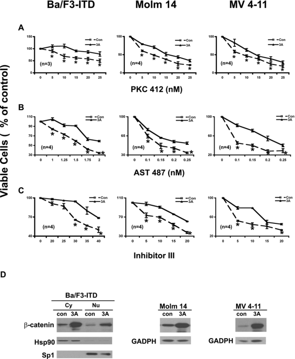
Figure 6.

Exogenous activation of the Wnt/β-catenin pathway by the Wnt3A ligand attenuates the potencies of the FLT3 kinase inhibitors on cell proliferation in either Ba/F3 cells ectopically expressing FLT3-ITD or cell lines derived from acute myeloid leukemia (AML) patient samples harboring FLT3-ITD mutations. (A) Effects of PKC412 on proliferation of the FLT3-ITD–expressing cells with and without the Wnt3A ligand stimulation. Ba/F3-FLT3-ITD cells were seeded at a density of 1.5 × 10⁵ cells/mL; Molm 14 and MV 4-11 cells were seeded at a density of 2 × 10⁵ cells/mL in the medium without IL3 but containing either 20% of 3A conditioned medium (3A) or 20% of L conditioned medium (Con) and treated with PKC412 at the concentrations indicated for 68 to 72 h, and then stained with trypan blue and counted under microscope. (B, C) Effects of AST 487 and inhibitor III on the proliferation of FLT3-ITD–expressing cells with and without the Wnt3A ligand stimulation. Analysis was carried out as in (A). (D) Expression levels of β-catenin after Wnt3A ligand stimulation. Ba/F3-FLT3-ITD cells were collected after incubation with either 20% Wnt3A conditioned medium or 20% of L conditioned medium (as control) for 1 h. The proteins of cytoplasmic portion and nuclear portion were separated and subjected to immunoblotting analysis using an antibody against β-catenin. The membrane was stripped and reprobed with Hsp 90 antibody as a marker for the cytoplasmic extraction; the membrane was stripped again and reprobed with Sp1 antibody as a marker for the nuclear extraction. Both Molm 14 and MV4-11 cells were treated with either 20% of the Wnt3A conditioned medium or 20% of the L conditioned medium for 3 h; the whole-cell lysis in NP-40 lysis buffer was then subjected to immunoblotting analysis using an antibody against β-catenin.
