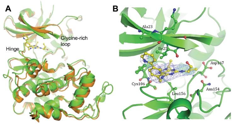
Figure 5.

Co-crystal structure of CR8 with CDK9/cyclin T. (A) (S)-CR8 bound to CDK9 (green) within the adenosine triphosphate (ATP) binding site in comparison to the apo kinase (orange, 3BLH). The 2 CDK/cyclin complexes were superimposed on their cyclin domains. (B) Close-up view of (S)-CR8 bound within the ATP binding pocket of CDK9/cyclin T. Hydrogen bonds are illustrated with dashed lines, and the final 2F0-Fc map contoured at 1σ is displayed as a blue mesh.
