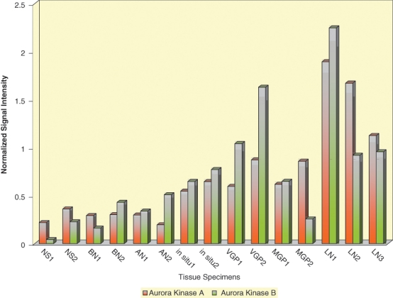
Figure 1.

Expression of Aurora kinases A and B in normal skin and nevus and melanoma tissue specimens subjected to whole-genome microarray analysis. Levels of Aurora kinase A (orange-colored bars) and Aurora kinase B (green-colored bars) expression in cryopreserved tissue samples representing normal human skin (NS1, NS2), benign nevi (BN1, BN2), atypical nevi (AN1, AN2), melanomas in situ (in situ1, in situ2), VGP melanomas (VGP1, VGP2), MGP melanomas (MGP1, MGP2), and melanoma-infiltrated lymph nodes (LN1, LN2, LN3).
