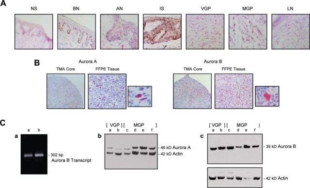
Figure 2.

Aurora kinase A and Aurora kinase B expression in cryopreserved and archival nevus and melanoma tissues and VGP and MGP melanoma cell lines. (A) Cryopreserved tissue sections, prepared from normal human skin (NS), a benign (BN) and an atypical nevus (AN), a melanoma in situ (IS), a VGP melanoma (VGP), an MGP melanoma (MGP), and a melanoma-infiltrated lymph node (LN), were probed with an antibody to Aurora kinase B. (B) Images of 2 representative tissue cores of a melanoma tissue microarray (TMA), probed with an antibody to Aurora kinase A and likewise an antibody to Aurora kinase B. Depicted in addition are 2 adjacent tissue sections, prepared from an FFPE MGP melanoma, that were stained by standard immunohistochemistry with an antibody to Aurora kinase A and likewise an antibody to Aurora kinase B. Next to each of the 2 tissue sections is an image, captured at higher magnification, which shows individual cells in the Aurora kinase antibody-probed FFPE MGP melanoma tissue. All tissue sections depicted (A and B) were counterstained with hematoxylin. (C) (a) RT-PCR analysis of Aurora kinase B mRNA expression in WM1158 (lane a) and WM983-B (lane b) MGP melanoma cell lines. (b) Immunoblot analysis of Aurora kinase A and (c) Aurora kinase B protein expression in VGP melanoma cell lines WM983-A (lane a) and WM98-2 (lane b) and in MGP melanoma cell lines WM373 (lane c), WM852 (lane d), WM983-B (lane e), and WM1158 (lane f). The immunoblots were probed with an antibody to β-actin serving as loading control.
