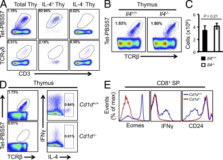
Figure 4.
iNKT cells are the source of IL-4 in the thymus. (A) Representation of iNKT cells and γδ T cells in gates of total thymocytes, IL-4+ thymocytes, and IL-4− thymocytes from wild-type BALB/c mice after stimulation with ionomycin and PMA in the presence of brefeldin A for 4 h. Tet, CD1d tetramer. (B) iNKT cells from the thymi of wild-type or IL4−/− BALB/c mice as measured by Tetramer-PB557 binding. (C) Absolute number of iNKT cells from the thymi of wild-type or IL4−/− BALB/c mice. Error bars are SDs. (D) Thymocytes from wild-type and Cd1d−/− BALB/c mice stained with Tetramer-PB557, anti–IFN-γ, anti–IL-4, and anti-TCRβ after stimulation as in A. (E) Expression of Eomes, IFN-γ, and CD24 on CD8+ SP cells from wild-type and Cd1d−/− BALB/c mice. Data are representative of three independent experiments with three mice in each genotype group (A–E).

