Figure 5.
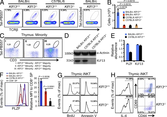
KLF13 regulates iNKT cells in BALB/c mice. (A) iNKT cells (Tet-PBS57+) in thymi of wild-type and Klf13−/− on BALB/c or C57BL/6 backgrounds. (B) Absolute number of thymic iNKT cells in indicated mice. (C) Minority iNKT cells (Tet-PBS57+) from lethally irradiated mice adoptively transferred with unequally mixed bone marrow cells, as indicated. (D) Expression of indicated genes in DP thymocytes from indicated mice measured by Western blot. (E) Abundance of indicated genes in FACS-sorted iNKT cells from indicated mice measured by quantitative real-time PCR. Error bars are generated from real-time PCR triplicates from the same RNA sample. (F) PLZF expression in thymic iNKT cells from the indicated mice (left). The mean fluorescence intensity (MFI) of PLZF in iNKT cells relative to CD8+ SP cells was determined using three mice for each point (right). (G) BrdU uptake and Annexin V binding to thymic iNKT cells from wild-type and Klf13−/− BALB/c mice. (H) Expression of IL-4, CD69, and CD44 by thymic iNKT cells from wild-type and KLF13−/− BALB/c mice. IL-4 was analyzed 4 h after stimulation with Ionomycin and PMA in the presence of brefeldin A. Data are representative of three independent experiments (A–H) with three mice in each genotype group (A–C and F–H). Error bars are SDs (B and F) or SDs from triplicate (E).

