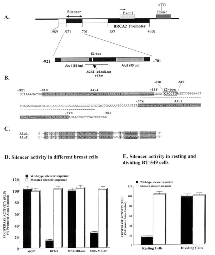
Fig. 1. Structure and function of human BRCA2 gene silencer.
A, a map of the promoter and the silencer at the upstream of the human BRCA2 gene. The putative ACR1 binding site at the silencer is indicated by a line. B, the nucleic acid sequence of the silencer showing the AluI, the AluII, and the E2-box. The 55-bp direct Alu repeats are in gray. The E2-box sequence (5′-CACCTG-3′) is boldfaced and boxed. The putative ACR1 binding site at the silencer is underscored. C, alignments of nucleotide sequences of the AluI/AluII. D, a Dual luciferase assay (Promega) to evaluate the function of the wild-type and E2-box-mutated silencer. Data are presented as mean (n = 12) ± S.E. The differences between the luciferase activities with the wild-type silencer sequence and the E2-box-mutated silencer sequence containing construct in BT-549 cells and in MDA-MB-231 cells are statistically significant (p < 0.0001). RLU, relative light units. E, activity of the silencer in the non-dividing (resting) and the dividing BT-549 cells. The differences between the luciferase activities with the wild-type silencer sequence and the E2-box-mutated silencer sequence-containing construct is statistically significant (p < 0.0005). The silencer DNA was mutated at the E2-box from 5′-CACCTG-3′ to 5′-AACCTA-3′.

