
Fig 2.
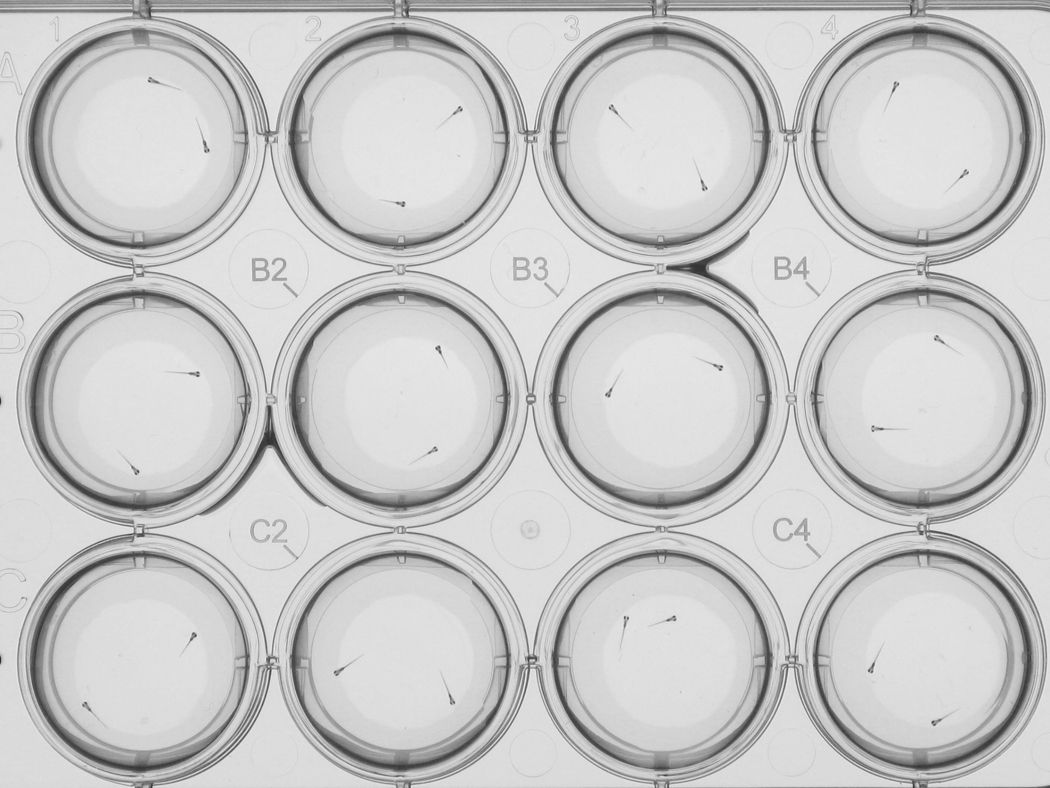
Zebrafish larvae imaged in a 12-well plate at 7 days post-fertilization. The larvae were imaged using a high-resolution imaging system that allows for the automated analysis of larval locations and orientations. For a description of the imaging system see (Colwill and Creton, 2010; Creton, 2009).