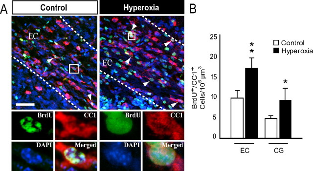
Figure 5.
Increased oligodendrogenesis after hyperoxia exposure throughout the immature white matter. Six-day-old (P6) mouse pups were exposed to normoxia (room air, 21% oxygen) or hyperoxia (80% oxygen) for 48 h. Animals in both experimental groups were kept in room air from P8 to P12. BrdU injections were started immediately at the end of exposure and were given every 24 h, and newly generated oligodendrocytes were labeled by immunofluorescence for CC1 and BrdU at P12. A, Confocal images of CC1+BrdU+ oligodendrocytes in control versus hyperoxia-exposed mice at P12. Arrowheads indicate triple immunolabeling for CC1, BrdU, and DAPI. Scale bar, 50 μm. B, Total numbers of newly formed CC1+BrdU+ oligodendrocytes in the EC and CG after cummulative BrdU injections from P8 to P12. Data are shown as mean ± SD (n = 3–5 brains for each group, using an unpaired t test comparing control vs hyperoxia; *p < 0.05; **p < 0.025).

