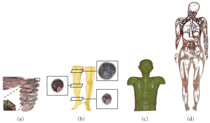
Figure 2.
Thick fascia connective tissues in VCH images were marked (green) (a) and their 3 D structures were rendered (b, c). When fascia connective tissues of the whole body, including thick and thin fascia tissues, were marked and their 3 D structures were reconstructed, a complete fascia network was observed; (d see reference no. [19]). All of the human organs and tissues were observed to be coated with connective tissues, and the connective tissues extended into the organs to form septa within the organs.

