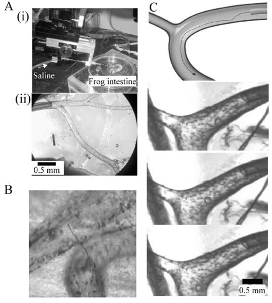
Fig. 6.

Assessment of nanowire electrode in frog mesentery artery.
(A) Experimental setup. (i) Microcatheter and frog intestine. (ii) Polyimide microcatheter (ϕ300 μm) is inserted into the mesentery artery. (B) Thick Pt wire of ϕ10 μm penetrates the blood vessel. (C) Fine Pt wire of ϕ1 μm is able to pass through the branch with a radius of curvature of about 700 μm in the 5 mm/s saline flow. The top panel shows a schematic diagram of the experiments. The bottom panels are time-series single video frames. Circle indicates location of electrode.
