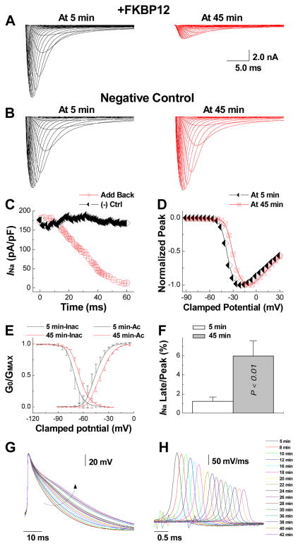
Figure 8.
Direct delivery of exogenous FKBP12 protein into FKBP12f/f/αMyHC-Cre+ ventricular cardiomyocytes replicates the effects of chronic FKBP12 overepxression on INa density and gating. A, Representative INa traces obtained from an FKBP12f/f/αMyHC-Cre+ ventricular cardiomyocyte at 5 and 45 minutes into continuous dialysis with purified recombinant FKBP12 protein (1 μg/μl). B, INa remained unchanged in an FKBP12f/f/αMyHC-Cre+ ventricular cardiomyocyte dialyzed with FKBP12-free pipette solution. C, Temporal changes in peak INa during continuous internal dialysis with FKBP12-containing and FKBP12-free, pipette solution, respectively. D, Normalized peak INa-V relationship at 5 and 45 minutes into FKBP12 dialysis. Internal perfusion of an FKBP12f/f/αMyHC-Cre+ ventricular cardiomyocyte with FKBP12-free solution did not alter voltage-dependence of INa activation (not shown). E, Changes in voltage-dependence of INa activation and inactivation following FKBP12 application. F, The late INa/peak INa ratio is significantly increased following 45 minutes of FKBP12 dialysis. G, Representative action potential recordings over the course of FKBP12 dialysis. The arrow indicates the gradual prolongation of APD. H, Traces of dV/dt for the same action potentials shown in G. Traces were shifted along the time axis for display purposes.

