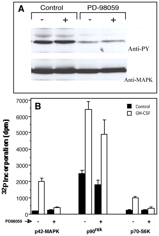
Figure 1. Inhibition of p70S6K activity by the MEK inhibitor PD-98059.
(A) Neutrophil phenotype-expressing cells dHL-60 (HL-60 cells continuously cultured in the presence of 1.25% DMSO for three days) were incubated with 25 μM PD-98059 for 30 minutes, followed by a short (5 min.) incubation with (+) or without (−) 270 pM GM-CSF and cell lysates were generated. Resulting proteins were analyzed by Western blotting with anti-phosphotyrosine antibodies (PY) and subsequently by anti-MAPK to confirm that equal amount of protein was loaded in each lane. (B) dHL-60 cells were incubated with 25 μM PD-98059 for 30 minutes and cell lysates were generated as in (A). These were split into three sets that were used for in vitro kinase assays against MAPK peptide substrate APRTPGGRR (left group of bars); p90rsk peptide substrate RRLSSLRA (middle group of bars); and p70S6K peptide substrate KKRNRTLTK (right group of bars). Results are the mean ± SE of four independent experiments performed in duplicate.

