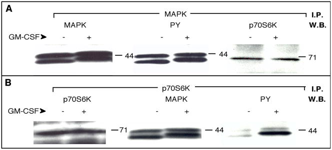
Figure 2. Co-immunoprecipitation of p70S6K and MAPK proteins.
(A) Neutrophils (1×10⁷ cells/ml) were incubated in the presence (+) or the absence (−) of 300 pM GM-CSF for 5 minutes. Lysates were immunoprecipitated (I.P.) with anti-MAPK (ERK1+2) antibodies and Western blots (W.B.) derived from immunocomplex beads were subjected to probing with anti-MAPK (left panel) followed by stripping of the antibody and re-probing with anti-PY (middle panel). Shown in both cases are the regions of the blot around the 44 kDa protein marker. Finally, the blot was stripped and re-probed with anti-p70S6K antibodies (right panel, showing the region of the blot around the 71 kDa marker). (B) Neutrophils immunoprecipitated with anti-p70S6K (C-18) antibodies followed by Western blotting with the same antibody (left panel, showing the region of the blot around the 71 kDa protein marker) and subsequent re-probing with anti-MAPK (middle panel, showing the 44-kDa region of the blot). Finally, the blot was probed again with anti-PY (PY20) (right panel, shown again is the region ~ 44 kDa). Results are typical among three other experiments.

