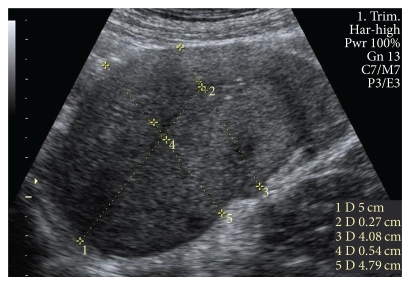
Figure 1.
Representative ultrasonographic image illustrating how the AP diameter of the uterus and the cavity were measured. (1) measured 5 cm from the fundus; (2) AP diameter of the cavity 5 cm from the fundus; (3) AP diameter of the uterus 5 cm from the funus; (4) maximum AP diameter of the cavity; (5) maximum AP diameter of the uterus.

