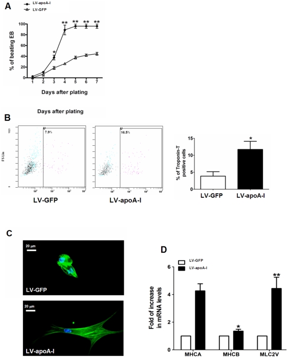
Figure 2. Effect of apoA-I gene transfer on generation of beating embryoid body and cardiac cells.
(A) Empty construct and apoA-I-transduced mouse ESCs were differentiated using the conventional “hanging-drop” method, and the resultant embryoid bodies (EBs) were plated onto gelatin coated plates. The occurrence of beating areas within the EBs was observed and counted for 8 days starting from the day of plating. (B) Percentage of ESC-derived cardiomyocytes (troponin-T positive cells) on day 8 as determined by flow cytometry. (C) Individual cardiomyocytes were isolated from the beating area of the EBs and identified with immunnohistochemistry using antibody specific to the cardiac troponin-T. (D) Cardiac maker gene expression in the embryoid bodies derived from empty construct- and apoA-I-transduced ESCs as revealed by real-time quantitative PCR analysis. MHCA: α-myosin heavy chain; MHCB: β-myosin heavy chain; MLC2V: myosin light chain 2 ventricular transcripts. Data shown as mean ± SEM from at least 3 independent experiments, n = 3–5, *p<0.05; **p<0.005.

