
Fig 4.
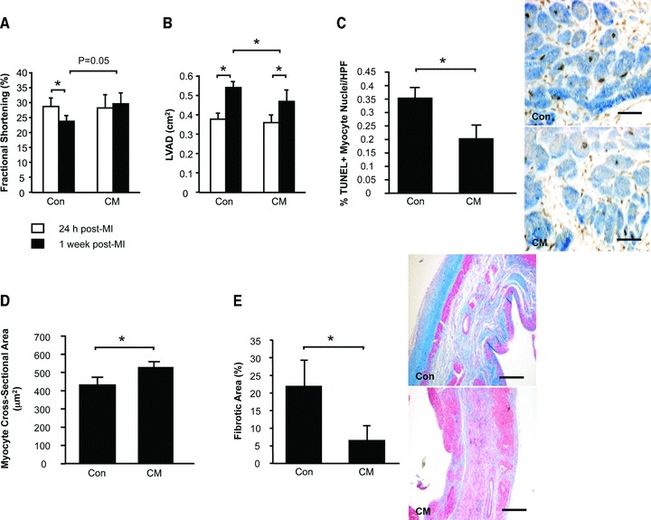
Intramyocardial injection of CM after MI attenuates left ventricular dysfunction and remodelling. Echocardiography was performed to assess FS (A) and LVAD (B). Myocardial sections were stained to count TUNEL+ apoptotic myocytes (C). Masson’s Trichrome-stained sections were assessed for myocyte cross-sectional areas (D) and myocardial fibrosis (E). Data are presented as mean +/− S.E.M. N = 10-12/group.