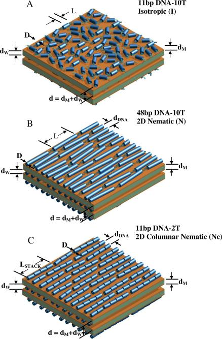
Figure 1.
Schematic drawings of the distinct packing phases of sDNA rods (all of which contain non-sticky overhangs) in cationic liposome–short DNA (CL–sDNA) complexes. The complexes are multilamellar assemblies of alternating cationic lipid bilayers of thickness dM and water layers of thickness dW. The water layer contains a monolayer of sDNA molecules. The multilamellar unit cell dimension is d = dW + dM. (A) The isotropic phase with short-range positional and orientational order, as observed for 11bp DNA-10T (11 bp DNA core with a 10-T non-sticky overhang at each end), with small shape anisotropy (length/width = L/D ≈ 1.9). (B) The nematic liquid crystal phase, as observed for 48 bp DNA rods with non-sticky ends (L/D ≈ 8.16). Formation of this phase by sDNA rods with sufficiently anisotropic shape is consistent with Onsager's model of LC ordering of anisotropic rods. For 5-T and 2-T non-sticky ends, some transient dimers may form, indicating the onset of sDNA end-to-end interactions. All rods are the same length (apparently shorter rods are shown cut off at the edges). (C) A new type of 2D columnar nematic phase, as observed for 11bp DNA-2T rods. The onset of strong DNA end-to-end interactions for 11 bp DNA with very short (2 T) non-sticky overhangs leads to the formation of a distribution of 1D stacks of sDNA rods, which become the building blocks of the nematic phase. The 1D stacks have an average size of ≈4 rods, corresponding to an effective length to width ratio L/D ≈ 7.3). In parts B and C, dDNA corresponds to the average interhelical spacing. The “Onsager nematic” (N) (part B) and columnar nematic (NC) (part C) phases exhibit long-range orientation and short-range position order. The drawings are not meant to imply that the orientation of sDNA rods is correlated between layers.

