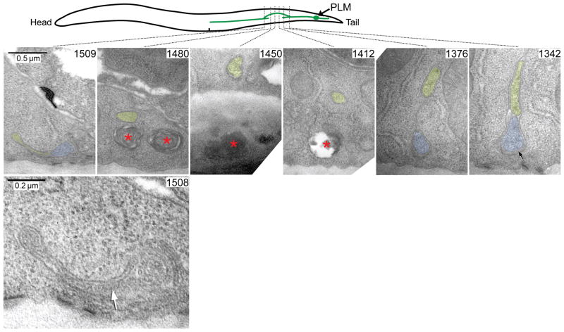
Figure 2.
Analysis of axonal reconnection with transmission electron microscopy. Serial thin sections of a wild-type animal 24 hrs post-axotomy are shown, along with a scheme to demonstrate how the PLM axon regrew around the laser damage zone. High magnification micrographs show the original axon (blue) and the new process (yellow) as it branched off from the original axon (section #1342), travelled inwards from the body wall (section #1376), traversed the damage site (section #1412 – #1480) and then fused as a very thin process (yellow) to the distal axon segment (blue) at section #1509. The fusion site is shown at higher power in section #1508, where the white arrow indicates the fusion zone. Local membrane whorls and voids (red asterisks) caused by collateral laser damage in the hypodermis can be seen in sections #1412, #1450 and #1480. Extracellular space below the PLM axon in section #1342 is swollen by mantle protein (black arrow), a characteristic feature of the mechanosensory neurons. Scale bars: 0.5 μm and 0.2 μm in sections #1509 and #1508, respectively.

