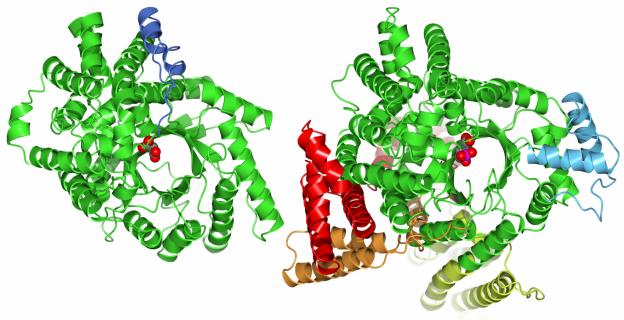
FIGURE 1.
The C. perfringens PepcA fold (left) and the E. coli Pepc fold viewed from above the active-site at the C-terminal end of the β-barrel. Insertions in the folds are indicated via the color scheme (compare to Figure 3). The insertions in the E. coli fold are at the N-terminus (red), after β-strands β1 (orange), β2 (yellow) and β3 (light blue). The helix-turn-helix insertion in the C. perfringens fold follows residue 360 (blue). Malonate is bound in the active-site of PepcA; a PEP analog is bound in the active-site of Pepc.

