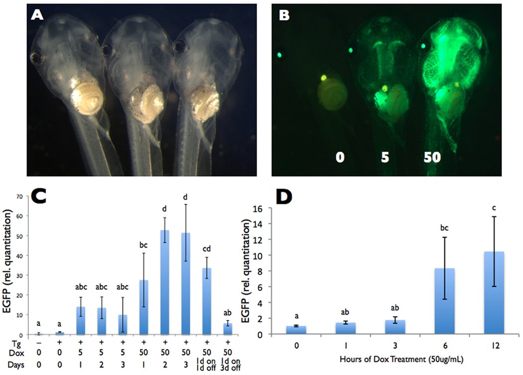
Figure 3.
Dose-dependent Dox-induced GFP expression. A and B) Five-day old offspring from founder ♂8 transgenic for pDPCrtTA-TREG-HS4 were treated with 0, 5, or 50 ug/mL Dox for one day and shown under A) brightfield and B) green fluorescence microscopy. In the absence of Dox, GFP is not visible in the body. In the presence of Dox, GFP is induced to a higher level with 50 compared to 5 ug/mL. A representative tadpole from the highest brightness class within each Dox treatment is shown. C and D) For a time course of dose-dependent GFP mRNA expression, GFP mRNA levels in the tail relative to the housekeeping gene rpL8 were measured (C) at 0, 1, 2, and 3 days of 5 and 50 ug/mL Dox treatment and (D) at 0, 1, 3, 6, and 12 hrs. of 50 ug/mL Dox treatment in 5- to 8-day old offspring from founder ♂8 transgenic for pDPCrtTA-TREG-HS4 using real-time reverse transcriptase PCR. Non-transgenic tadpoles were included as controls. C. Sample size was n=3, and no tadpoles were pooled. To avoid variation in the results due to variation in GFP expression amongs siblings in founder ♂8 (seen in Fig. 2C), tadpoles from the highest brightness class were used for all time points (except Day 0), done by selecting similarly bright individuals from a surplus of Dox-treated tadpoles after 24 hours. D. Because tadoles could not be preselected based on GFP brightness prior to tissue harvesting, we used a sample size of n=3 and pooled four tadpoles per sample to decrease the chances of obtaining unrepresentative results, given the expected levels of variability in GFP expression among siblings in the same treatment. Letters above the bars represent significance groups based on ANOVA and Bonferroni post-hoc pairwise comparisons, α = 0.05.

