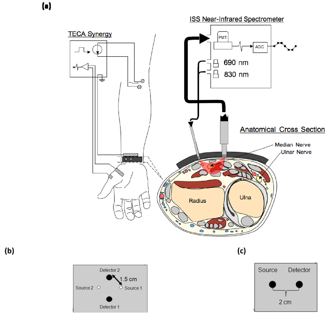
Figure 2.
Experimental setup for the human studies. (a) Configuration of the Ag/AgCl electrodes and optical probe while making CMAP measurements of APB during stimulation of the median nerve at the elbow. The anatomical cross section illustrates the location of the median and ulnar nerves in the region of optical interrogation. (b) Probe geometry used for the vascular occlusion protocol. (c) Probe geometry for the spectral dependence protocol.

