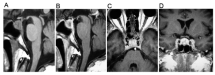
Figure 1.
A cranial magnetic resonance imaging showed an intrasellar mass. (A) A sagittal T1-weighted image revealed a sellar lesion with hypointense areas. (B) A gadolinium-enhanced sagittal image showed an enhancing lesion with a hypointense center. Axial (C) and coronal (D) images demonstrated parasellar extension toward the left cavernous sinus.

