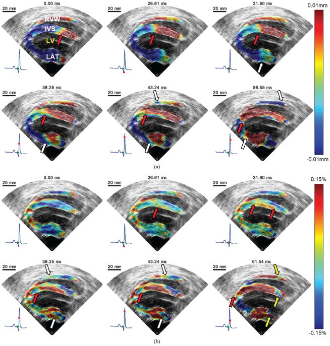
Fig. 4.
EWI during sinus rhythm in the parasternal four-chamber view in the normal ventricles. RVW, IVS, LV, and LAT, respectively, denote right ventricle wall, interventricular septum, left ventricle cavity and free wall (lateral wall). (a) Incremental displacement images. Displacements away and towards the transducer are depicted in red and blue, respectively. A wave traveling from base to apex is clearly visible in the septum. (b) Incremental strain images. Approximately 30 ms after the onset of the QRS, excitation of the upper endocardium of the left ventricle begins. It then propagates both towards the base and the apex (red arrows). Mechanical activation also appears in the lateral wall and travels mostly towards the base. 40 ms after the onset of the QRS, the right ventricular wall begins to thicken. 60 ms after the onset of the QRS, the myocardium is still not completely mechanically activated; thinning of portions of the base are still visible (yellow arrows). At 61.54 ms, negative incremental strains are observed in the apical region. This is most likely due to the fact that in this region, the axial direction contains a large projection of the longitudinal strain component.

