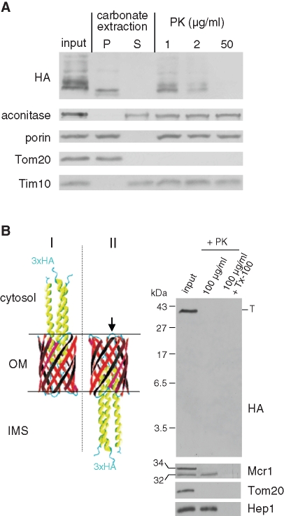
FIGURE 4:
YadA-MA is integrated into the mitochondrial outer membrane in the correct topology. (A) Mitochondria isolated from cells expressing YadA-MA were loaded directly on SDS–PAGE gel (input), or were first subjected to carbonate extraction and then centrifuged to discriminate between membrane proteins in the pellet (P) and soluble proteins in the supernatant (S). Additional aliquots of mitochondria were treated with the indicated amounts of PK. Proteins were analyzed by SDS–PAGE and immunodecorated with antibodies against the indicated proteins: aconitase, a mitochondrial matrix protein; porin, a protein embedded in the outer membrane; Tom20, an outer membrane protein exposed to the cytosol; Tim10, a soluble IMS protein. (B) Left, models of two putative conformations of HA-tagged YadA-MA in the mitochondrial outer membrane. The native (I) and the upside-down conformation (II) are displayed with an arrow pointing putative PK-sensitive loops for the upside-down conformation. Right, PK protection assay of mitochondria isolated from yeast cells expressing HA-tagged YadA-MA. Mitochondria were left untreated (input) or were treated with PK in the absence or presence of Triton X-100 (Tx-100). The samples were analyzed by SDS–PAGE on a gel optimized for detection of small polypeptides followed by immunodecoration with antibodies against HA, Tom20 (outer membrane), Hep1 (matrix), and Mcr1 (outer membrane and IMS). The latter protein has two isoforms: a 34 kDa form exposed at the outer membrane and a 32 kDa form in the IMS. Molecular mass markers are indicated on the left. The trimeric form of YadA-MA is indicated to the right with T.

